What Is the Biological Fate of Asbestos?
Upon completion of this section, you will be able to
- Identify where asbestos fibers are most likely to be retained in the body.
Inhaled asbestos fibers are deposited in the upper and lower respiratory tracts. The durability and retention of the fibers in lung tissue and elsewhere in the body may lead to a risk of disease.
Asbestos particles occur in many sizes in inhaled air. The largest size asbestos particles tend to deposit on the nasal mucosa or the oropharynx and are sneezed out or swallowed never reaching the lungs [NIOSH 2011a].
Some of the smaller inhaled asbestos fibers are deposited on the surface of the larger airways where some of them are cleared by mucociliary transport and swallowing. Other smaller fibers are deposited further down in the lung, especially in the bifurcations of the tracheobronchial tree, and some are deposited in the alveolar sacs [Broaddus 2001].
There has been much scientific discussion about how fiber size affects the pathogenicity of asbestos. It is believed that the dimensions of the asbestos fiber determines how far into the lungs it is likely to be deposited and how quickly it is cleared. Wide fibers (diameter greater than 2 to 5 microns) tend to be deposited in the upper respiratory tract and cleared. Some recent papers have shown cumulative fiber exposure to be an important risk factor for the development of asbestosis [Larson et al. 2010a].
However, it is important to remember that asbestos fibers of all lengths cannot be excluded as contributors to asbestos-related diseases [Dodson et al. 2003].
Once deposited in the lungs, asbestos fibers are subject to several lung defenses.
- The most important means of removal of insoluble asbestos particles deposited in respiratory tract airways is by mucociliary clearance. This cleared material is usually swallowed and enters the gastrointestinal tract; however, the asbestos coughed out in sputum is eliminated from the body [NIOSH 2011a].
- The most important means of removal of insoluble asbestos particles deposited in the alveoli involves alveolar macrophages. These specialized cells generally function to clear particles depositing in the alveoli by enveloping them in a process called phagocytosis and then moving to the airways, where the particle-containing macrophages are transported out of the lungs via mucociliary clearance.
In some cases, insoluble particles are not easily cleared from the alveoli by alveolar macrophages.
- Some (e.g., crystalline silica) particles are highly reactive and toxic to the macrophage cells, which die before they can clear the engulfed particles.
- Under “overload” conditions so many particles are inhaled that they overwhelm the ability of alveolar macrophages to clear them from the alveoli.
- With increasing particle dimension greater than about 15 microns, phagocytosis by macrophages becomes increasingly ineffective. The inability of macrophages to engulf such particles results in “frustrated phagocytosis” [NIOSH 2011a].
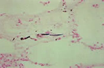
Some asbestos fibers deposited in alveoli make their way to the lymphatic system of the lungs, which helps clears fibers from the lungs [Dodson et al. 2003]. Still, many asbestos fibers are retained in lung tissue for many years. The total burden of residual fibers in the lungs depend not only the size of the fibers but the amount of fibers inhaled from the environment [Dodson et al. 2003].

Figure 4. Asbestos fiber retained in lung tissue
From the lungs, some asbestos fibers (mainly short fibers) can migrate to pleural and peritoneal spaces, especially following patterns of lymphatic drainage [Broaddus 2001]. Their presence in the peritoneum is more likely if there is a high fiber burden in the lungs.
How rapidly the body’s defenses can clear asbestos fibers from the lungs depends in part on the type of asbestos. While all types of fibers are retained for many years in the lungs [ATSDR 2001a], amphibole fibers are degraded more slowly than serpentine fibers of the same dimensions [Hillerdal 1999].
Most ingested asbestos fibers pass through the gastrointestinal tract unchanged and are cleared in the feces. A few ingested fibers pass through the walls of the gastrointestinal tract. Some of these fibers stay in the peritoneal cavity. Others move into the bloodstream and into the kidneys, where some are eliminated unchanged in the urine.
- Some inhaled asbestos fibers reach the lungs, where they become lodged in lung tissue, especially at bifurcations and in the lower lung fields.
- Some fibers move to pleural or peritoneal spaces or the mesothelium.
- The half-lives of asbestos fibers vary, but some are retained in the lungs for many years.
- Ingested asbestos fibers are usually eliminated from the body.